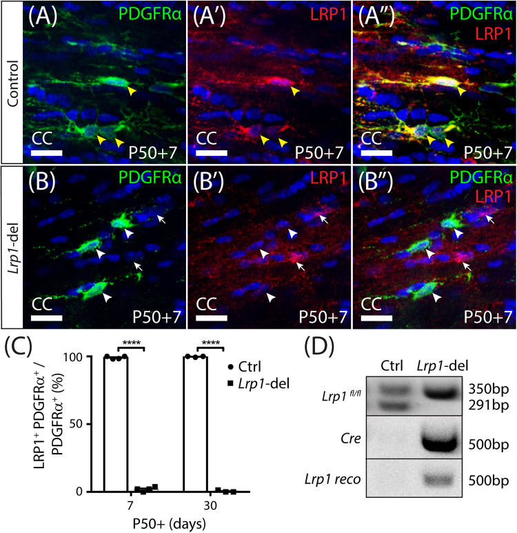
FIGURE 1.
Lrp1 can be conditionally deleted from adult OPCs at high efficiency. Coronal brain sections from P57+7 and P57+30 control (Pdgfrα-CreERTM) and Lrp1-deleted (Pdgfrα-CreERTM :: Lrp1fl/fl) mice were immunolabeled to detect OPCs (PDGFRα, green), LRP1 (red), and cell nuclei (Hoechst 33342, blue). (A–A″) Compressed z-stack confocal image of LRP1+ OPCs (solid yellow arrow heads) in the corpus callosum (CC) of a P50+7 control mouse. (B–B″) Compressed z-stack confocal image of LRP1-neg OPCs (solid white arrow heads) in the CC of a P50+7 Lrp1-deleted mouse. White arrows indicate PDGFRα-neg cells that remain LRP1+ in the Lrp1-deleted mice. (C) The proportion (%) of PDGFRα+ OPCs that express LRP1 in P50+7 and P50+30 control and Lrp1-deleted mice [mean ± SD for n ≥ 3 mice per genotype per time-point; 2-way ANOVA: Genotype F (1,10) = 2.8, p < 0.0001; Time F (1,10) = 0.52, p = 0.5; Interaction F (1,10) = 3.44, p = 0.09]. Bonferroni multiple comparisons ****p ≤ 0.0001. (D) PCR amplification of genomic DNA extracted from the brain of a P50+7 control (Lrp1fl/+) and Lrp1-deleted (Pdgfrα-CreERTM :: Lrp1fl/fl) mouse indicates that recombination, producing the Lrp1 reco band, only occurs in Lrp1-deleted mice. Scale bars represent 17 μm.

