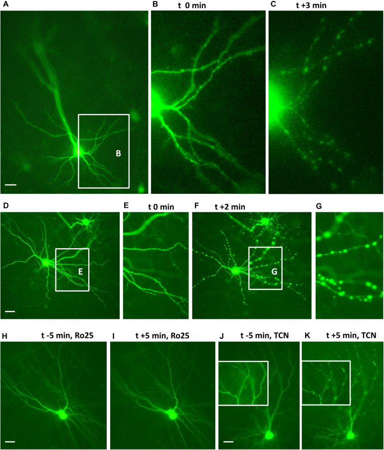
FIGURE 1.
Endogenous GluN2 receptors are functional on DIV 10 neurons. Cultures with live EGFP-expressing neurons were challenged with 50 μM NMDA to evoke dendritic beading. (A–C) Pyramidal neuron before and 3 min (t + 3 min) after application of NMDA. The inset in (A) shows basal dendrites which are seen at higher magnification in (B,C). (D–G): Interneuron before (D,E) and 2 min after NMDA application (F,G). The insets in (D,F) show the dendrites which are at higher magnification in (E,G), respectively. (H,I): Interneuron protected from the NMDA-induced dendritic beading by preincubation with 5 μM Ro25-6981. (J,K) Pyramidal cell not protected from the NMDA-induced dendritic beading by preincubation with 10 μM TCN201; insets show details of apical dendritic branches at higher magnification before and 5 min after application of NMDA. Pial surface is to the upper left for (A–I) and to the top for (J,K), Scales: 10 μm.

