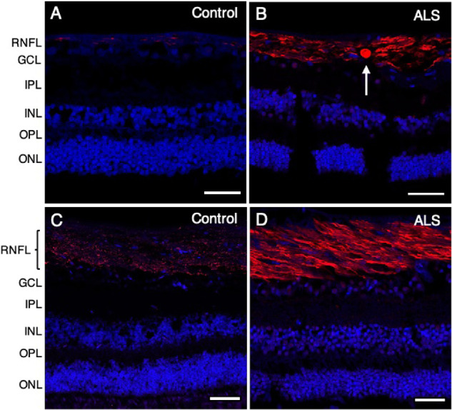
Figure 4.

Increased phosphorylated neurofilament in the peripheral and central retina of patients with ALS. Phosphorylated neurofilament is shown in red using SMI 31, with nuclei counterstained with DAPI in blue. P-NF–positive spheroids ranging from 8 to 15 µm in diameter were observed in the peripheral retina (B, arrow) but not in the central retina in patients with ALS (D). Compared with controls (A, C), a stronger P-NF signal was observed in patients with ALS (B, D) in both peripheral (B) and central (D) retinal regions. (A) Control (case 9); (B) ALS (case 7); (C) control (case 3); (D) ALS (case 5). Abbreviations: RNFL = retinal nerve fiber layer; GCL = ganglion cell layer; IPL = inner plexiform layer; INL = inner nuclear layer; OPL = outer plexiform layer; ONL = outer nuclear layer. Scale bar = 50 µm.
