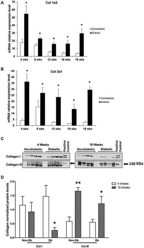
Figure 2.

Collagen gene and protein expression in diabetic and nondiabetic murine skin. (A) Relative gene expression for collagen Iα2 in skin samples from diabetic (n = 5) versus nondiabetic (n = 5) mice at 4 and 18 weeks of age. (B) Relative gene expression for collagen IIIα1 as measured in skin samples from diabetic (n = 5) versus nondiabetic (n = 5) mice at 4 and 18 weeks of age. (C) Collagen I and III (upper band; black arrows) levels as demonstrated by Western blots obtained from skin samples from age-matched, nondiabetic, and diabetic mice at 4 and 18 weeks of age. (D) Collagen I and III protein levels as quantified by Western blot. These findings are representative of five independent experiments. Data is presented as a mean + SEM for each cohort. Student’s t test was used to compare nondiabetic skin to diabetic skin at each time point, with *p < 0.05; **p < 0.001.
