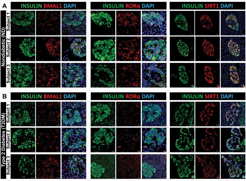
Figure 7.
BMAL1, RORα, and SIRT1 immunoreactivity is attenuated in human T2DM β-cells. Representative examples of pancreatic islets stained by immunofluorescence for BMAL1, RORα, and SIRT1 (red), insulin (green), and counterstained with nuclear marker DAPI (blue) imaged at × 20 (BMAL1 and RORα scale bars, 20 µm; SIRT1 scale bars, 50 µm) in human pancreatic tissue obtained from Mayo Clinic autopsy archives. Graph shows 3 representative examples of nondiabetic (ND; A) and T2DM (B) subjects. In total, pancreatic specimens from n = 8 (ND) and n = 7 T2DM subjects were obtained and examined.

