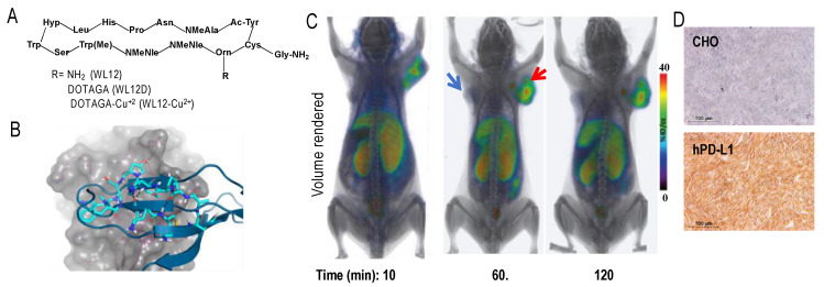
Figure 1.
WL12 binding interactions with PD-L1 overlap with those of PD-1. (A) Structural representation of WL12 and its analogs; (B) predicted binding mode of WL12 to PD-L1. WL12 forms a beta sheet-like structure in the groove of PD-L1. WL12 is shown in cyan. The surface representation of PD-L1 is shown in gray, with the ribbons and key side chains shown in magenta; WL12 mimics PD-1 binding to PD-L1. The structure of PD-1 is shown in teal. The two main interacting beta strands of PD-1 overlap well with the conformation adopted by WL12 bound to PD-L1. (C) NSG mice with hPD-L1 (red arrow) and CHO tumors (blue arrow) were administered intravenously with 150 μCi of [64Cu]WL12 and images were acquired at 10, 60, and 120 min after the injection of the radiotracer. 3D volume rendered images show specific accumulation of [64Cu]WL12 in hPD-L1 tumors. (D) PD-L1 IHC shows strong immunoreactivity (brown color) in hPD-L1 tumors (from [153]).

