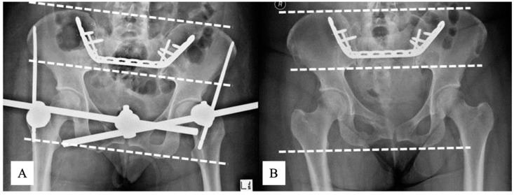
Figure 3.
AP X-rays of Patient 2 pelvis immediately after reduction and fixation (A) and at 104 weeks follow-up (B). In this patient, RSA measured a 1 cm fracture translation during healing. Note that there are no measurable vertical translations on plain radiographs, and that the iliac bones project relatively symmetric on each image. Note the slightly different projections of the pelvis between examinations. The dotted lines correspond to a horizontal plain. AP, antero-posterior; RSA, radiostereometric analysis;

