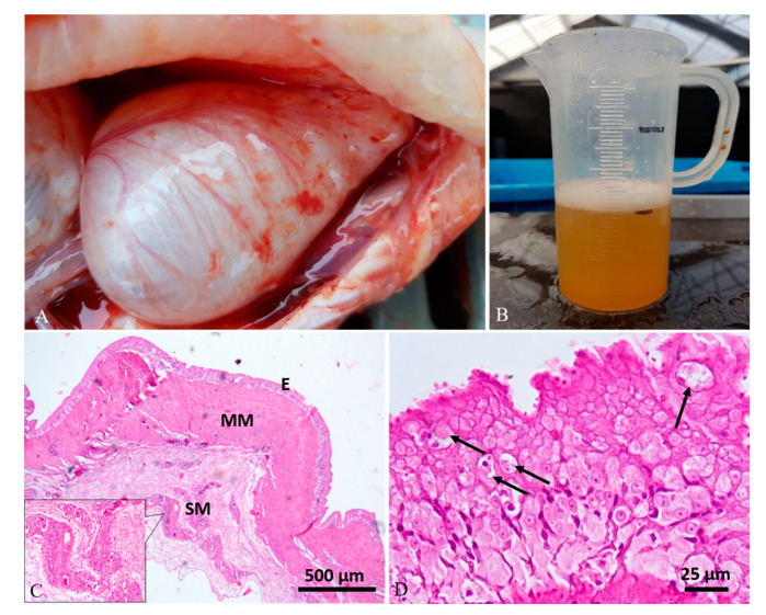
Figure 1.
Case 4#. (A) Swim bladder showing a severe enlargement of chambers and multifocal superficial hyperemic streaks. (B) A conspicuous amount of yellow clear fluid was aspirated from the swim bladder. (C) Histological analysis showed severe thickening of the muscularis mucosae (MM) and perivascular inflammatory-cell infiltration in the submucosa (SM) (inset; hematoxylin–eosin (H&E) stain). (D) The mucosa (E) showed a severe epithelial hyperplasia with the presence of intracytoplasmic eosinophilic round aggregates compatible with parasitic stages (arrows) (H&E stain).

