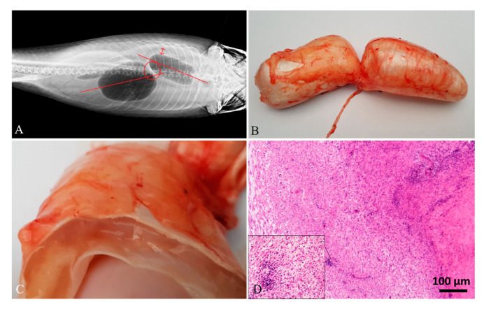
Figure 3.
Case 6#. (A) Radiographic image of swim bladder showing dislocation of the chambers. (B) Grossly, the size of the chambers appeared slightly enlarged. (C) When cut in a transversal section, abundant gelatinous material expanding the swim bladder layers was detected. (D) Histological analysis showed the muscularis mucosae expanded by abundant granulation tissue and a severe inflammatory cell infiltration, mainly composed of lymphocytes (inset; H&E stain).

