Figure 4.
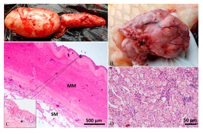
Case 1#. (A) Swim bladder showing a moderate enlargement of caudal chamber. (B) The fish had a large intracoelomic tumor, which caused the compression of the swim bladder. (C) Histological analysis of the swim bladder showed fibrous hyperplasia of the muscularis mucosae (MM), submucosal (SM) multifocal inflammatory cell infiltration, edema of lamina propria (asterisk in inset), and mucosal (E) hyperplasia with squamous metaplasia (inset; H&E stain). (D) Histology of the tumor revealed a gonadal germ-cell tumor (H&E stain).

