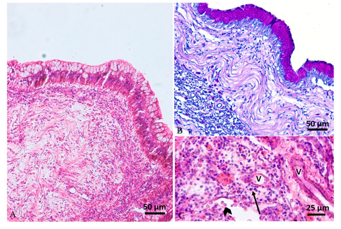
Figure 5.
Case 2#. (A) Histology of swim bladder showing mucosal hyperplasia with mucous metaplasia and a severe mixed inflammatory cell infiltration expanding the lamina propria and muscularis mucosae (H&E stain). (B) Periodic acid–Schiff (PAS) stain highlights mucous metaplasia of epithelium. (C) The inflammatory reaction, composed of mainly lymphocytes (arrow) and mast cells (arrowhead), was located mainly around vessels (V) (H&E stain).

