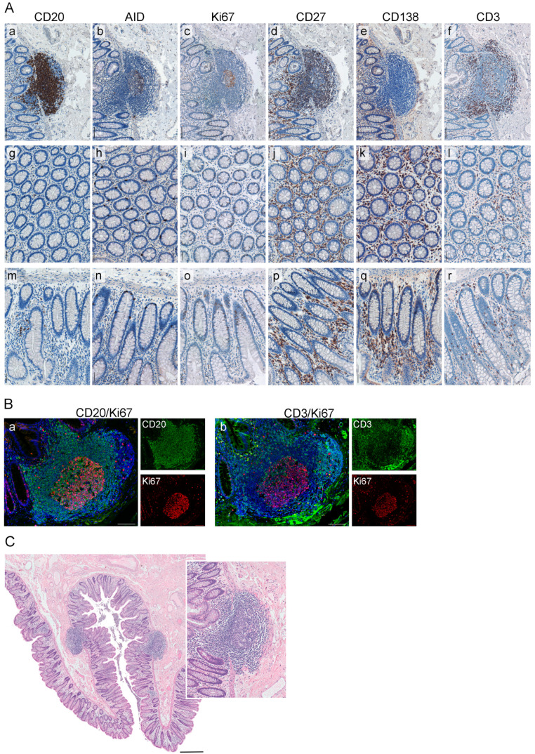
Figure 3.
Characterization of the immune phenotype of ILS within NT. (A) Shown are the representative images of an ILSs within NT, and the corresponding colonic mucosa whereat the FFPE tissue sections were stained by IHC for the markers CD20 (a, g, m), AID (b, h, n), Ki67 (c, i, o), CD27 (d, j, p), CD138 (e, k, q), and CD3 (f, l, r); color code: brown, the marker; blue, nuclear counterstaining with hematoxylin. Scale bar: 100 µm. (B) The proliferative status of B-cell and T-cell populations within ILSs was assessed by IF staining. Color code: (a) green, CD20; red, Ki67; blue, nuclear counterstaining by DAPI; (b) green, CD3; red, Ki67; blue, nuclear counterstaining by DAPI. Shown are images for individual channels and the merged images. Scale bar: 100 µm. (C) The presence and localization of compact round-shaped ILSs within normal colon mucosa of NT is visualized by HE staining. Scale bar: 500 µm. Insert: enlarged view of ILS with prominent GC area. FFPE, formalin-fixed paraffin-embedded; IHC, immunohistochemical; AID, activation-induced cytidine deaminase; NT, non-tumorous tissue; ILS, isolated lymphoid structure; IF, immunofluorescent, HE, hematoxylin and eosin; GC, germinal center; DAPI, 4′,6-diamidino-2-phenylindole.

