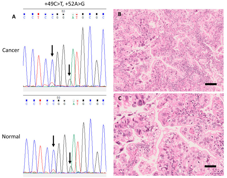
Figure 3.
Representative case with a germline single-nucleotide variant (SNV) in the rDNA promoter (case 1). (A) Electropherograms of germline SNVs +49C>T and +52A>G from corresponding cancer (upper panel) and normal (lower panel) tissue samples. (B) Representative histopathological images of this case (H&E stain) showing invasive mucinous carcinoma accompanied with papillary structure, and (C) nuclear pleomorphism and frequent mitoses. Scale bar, 50 μm.

