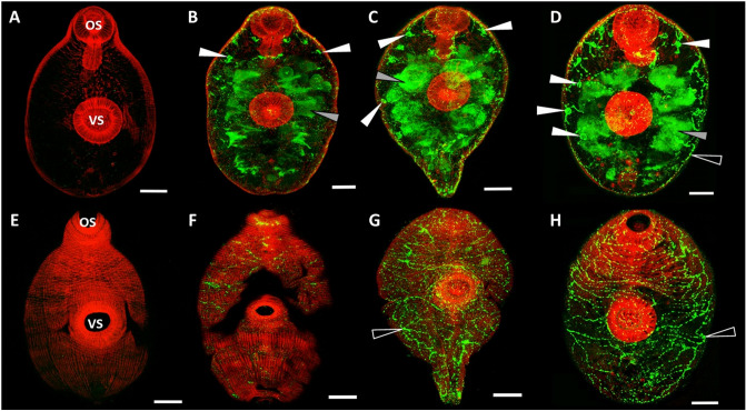
Figure 3.
Immunolocalization of F. hepatica KT in infective NEJs in the first 48 h of infection. (A–D) show a plane within the interior of the NEJ, whereas (E–H) show a plane at the surface of the same individual NEJs. Following excystment, NEJs were maintained in culture and sample parasites taken at 3 h post-excystment (B,F), 24 h post-excystment (C,G) and 48 h post-excystment (D,H). Parasites were probed with rabbit pre-immune serum (A,E) or anti-FhKT1 antibodies followed by FITC-labelled secondary antibodies. The presence of FhKT within F. hepatica-specific structures are indicated by arrows; parenchymal cell bodies, white arrows; NEJ digestive tract, grey arrows; network of channels below surface tegument, black arrows. All specimens were counter-stained with phalloidin-tetramethylrhodamine isothiocyanate (TRITC) to stain muscle tissue (red fluorescence) and provide structure. OS oral sucker, VS ventral sucker. Scale bars 20 μM.

