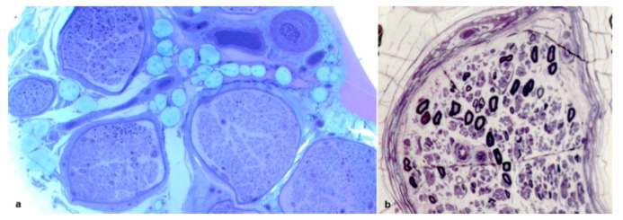
Figure 2.
Sural nerve biopsies from a 71-year-old Tyr78Phe (a) and from a 68-year-old Phe64Leu (b) hATTR patients. Semithin sections stained with toluidine blue. An inhomogeneous interfascicular fiber loss is evident (a): in two fascicles only isolated myelin fibers were present while other fascicles show an asymmetrical fiber loss. An inhomogeneous intrafascicular fiber loss is present (b).

