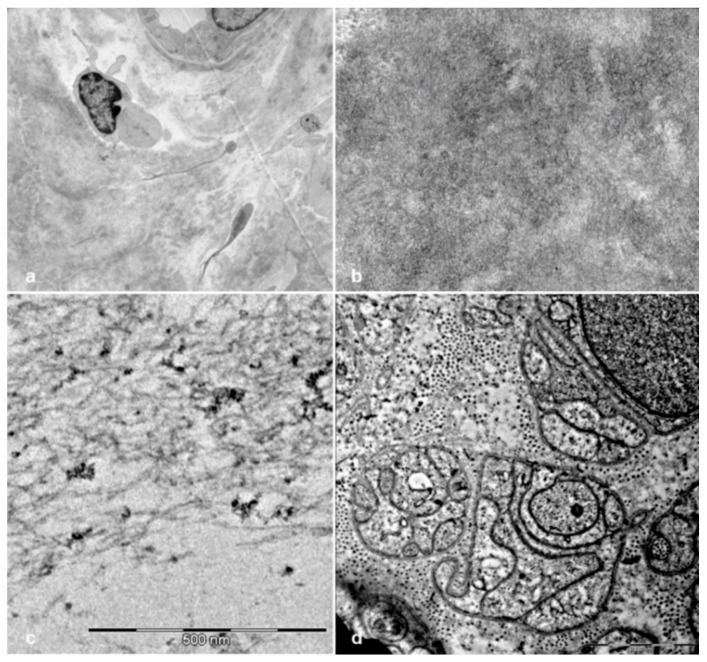
Figure 8.
Electron microscope examination of sural nerve biopsies from hATTR patients. Ultrathin sections stained with uranyl acetate and lead citrate. Amyloid deposits are evident in a biopsy from a 65-year-old Val30Met patient (a,b). Amyloid from a 73-year-old Val30Met patient shows typical fibrillar structure (c). Unmyelinated fibers from a 74-year-old Phe64Leu patient are reduced and replaced by collagen pockets (d).

