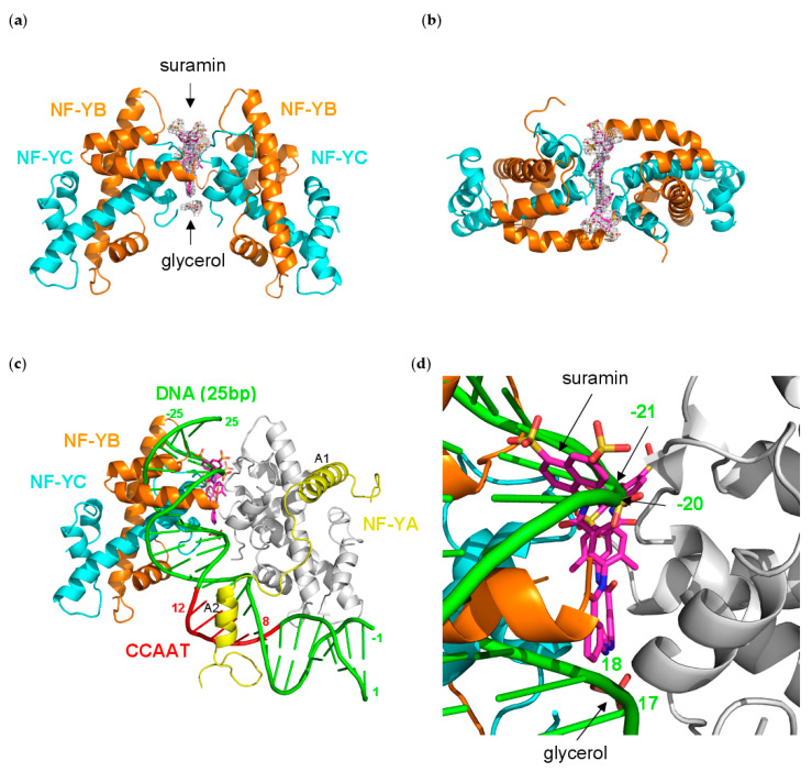
Figure 4.
Structure of the (NF-Yd)2–suramin complex; (a) ribbon diagram showing the bound suramin (magenta sticks) at the dimerization interface of two NF-Yd molecules (NF-YB in orange, and NF-YC in cyan), together with a glycerol molecule (from the cryoprotectant solution). A representative electron density map (grey net), contoured at 1.0 σ, is shown around the bound molecules; (b) top view of (NF-Yd)2–suramin complex; (c) Structural superposition of one NF-Yd molecule of the (NF-Yd)2–suramin complex with the DNA/NF-Y complex (NF-YB/NF-YC in grey, NF-YA in yellow, DNA in green) (PDB-code 4AWL). The CCAAT box is shown in red. The NF-YA A1 and A2 α-helices are labeled, and the DNA numbering is indicated. About 10 bp of the bound DNA superimpose with the second NF-Yd molecule of the (NF-Yd)2–suramin complex; (d) close-up of panel (c) showing that two sulfonic acid groups of suramin and the glycerol molecule approximately match the positions of two phosphate groups in both DNA strands of the DNA/NF-Y complex (−20, −21, and 17, 18, respectively).

