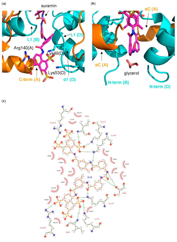
Figure 5.
Interactions of suramin in the NF-Yd structure; (a) close-up of the binding pocket of half-suramin at the NF-Yd dimerization interface. Secondary structure elements and protein chains are indicated. Color coding as in Figure 4; (b) glycerol-binding pocket relative to the suramin binding site. The glycerol molecule is shown in pink sticks; (c) schematic representation with LIGPLOT [60]. Polar contacts are depicted as broken lines and hydrophobic contacts are indicated by arcs with radiating spokes.

