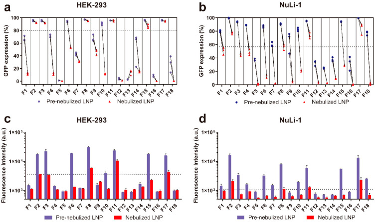
Figure 3.
In vitro intracellular protein expression in terms of percent GFP expression and fluorescence intensity of LNP formulations before and after nebulization in HEK-293 (a,c) and NuLi-1 cells (b,d). Figure 3a,b represent the percent GFP expression in HEK-293 and NuLi-1 cells, respectively. Figure 3c,d represent the GFP fluorescence intensity in HEK-293 and NuLi-1 cells, respectively.

