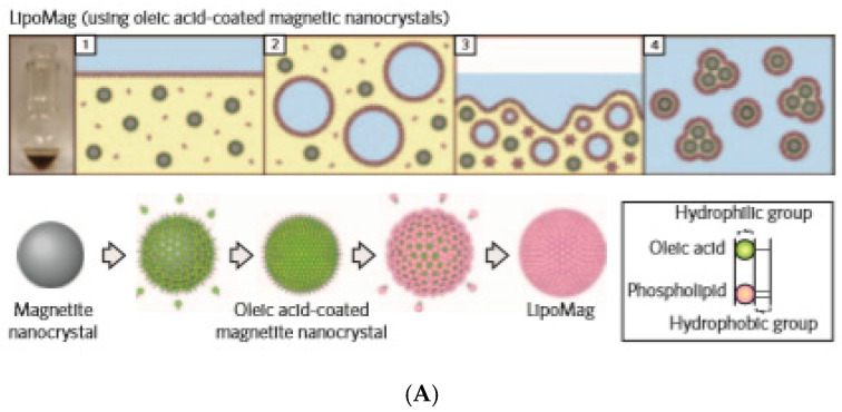
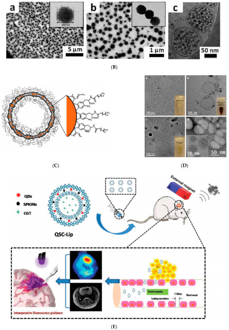
Figure 5.
(A) Schematic showing the preparation (upper) and assembly (lower) of LipoMag. Oleic acid-coated magnetic nanocrystal cores and the lipid shells form through hydrophobic interactions (reprinted by permission from Copyright Clearance Center: Springer Nature, Nature Nanotechnology, [158], Copyright 2009). (B) (a,b) TEM and (c) cryo-TEM micrographs of UMLs prepared by an REV process. At low magnification, a large number of dense vesicles are observed with diameters 200 nm in average. MNPs are trapped inside unilamellar vesicles (c) and dipole−dipole interaction can occur as exemplified by magnification (b). (Reprinted with permission from [64]. Copyright 2012 American Chemical Society). (C) Schematic of liposomes containing iron oxide NPs in their bilayer. NitroDOPA–palmityl-stabilized iron oxide NPs are embedded in liposome membranes consisting of PEGylated and unmodified lipids; (D) Liposomes functionalized with iron oxide NPs. Cryo-TEM images of DSPC liposomes containing 5 mol % PEG(2)–PE that (a) were unmodified and incorporated (b) oleic acid-coated and (c) palmityl–nitroDOPA stabilized small iron oxide NPs. Insets show photographs of the respective PbS-based liposome dispersions where the lipid concentration was kept constant at 5 mg/mL. A comparison between (a) and (c) reveals no significant change of the spherical shape of liposomes upon loading their membranes with small, individually stabilized, iron oxide NPs. However, agglomerated, oleic acid-stabilized NPs seem to significantly distort the liposome shape. (d) TEM image of trehalose-fixed DSPC liposomes containing palmityl–nitroDOPA stabilized small NPs in their membranes. Liposomes were fixed with trehalose and air-dried on a carbon-supported Cu TEM grid where the carbon film had 3.5 μm diameter holes. While the large image was taken in a hole that was spanned by trehalose, the inset was imaged on the carbon support. Individually stabilized NPs with core diameters <5.5 nm are associated with liposomes. No NPs with core diameters >5.5 nm are seen. The inset indicates a high NP density of liposomes that were collapsed on the carbon support upon drying in air. (Reprinted with permission from Amstad et al. 2011a. Copyright 2011 American Chemical Society). (E) Liposome-integrated multiple-imaging agents and therapeutic drug for glioma-targeted delivery under exogenous magnetic field to accurately localize glioma. CGT, cilengitaide; QDs, quantum dots and SPIONs, superparamagnetic iron oxide nanoparticles were initially dispersed in chloroform. (Republished with permission of John Wiley and Sons, from [163]; permission conveyed through Copyright Clearance Center, Inc.).


