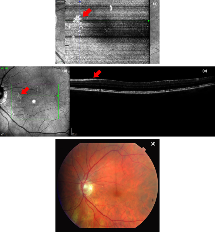
Figure 1.

(a) En face image of an 85‐year‐old control subject with high myopia and ARAM using a high‐density protocol. Patches of hyper‐reflectivity can be seen here in the same locations as the patchy, discrete, glistening lesions on the SLO image in (b). On the B‐scan (c), a hyper‐reflective line is seen at the ILM in the glittering area. In comparison, the ARAM cannot be seen in (d) a standard colour fundus photo of the same patient taken 15 min after OCT imaging.
