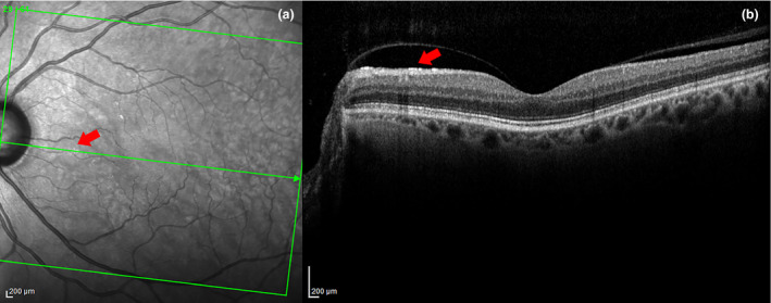
Figure 2.

In 2a, the arrow points to a very small patch of ARAM in the SLO image. In 2b, a hyper‐reflective layer is seen at the ILM in the B‐scan at the same location as the ARAM lesion on the SLO. A partial PVD with detachment in the area of the ARAM is also evident here. There are several other small patches of ARAM seen in (a) but could not be captured due to the sparse sampling of B‐scans.
