
Fig 2.
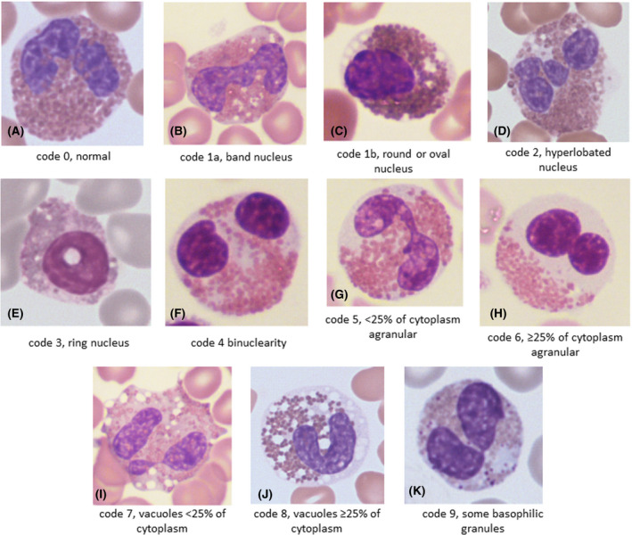
Representative examples of morphological features in patients with a myeloid neoplasm: (A) a normal eosinophil (code 0) from a patient with atypical chronic myeloid leukaemia evolving into acute myeloid leukaemia (AML); (B) a band eosinophil (code 1a) from a patient with FIP1L1‐PDGFRA; (C) an eosinophil with an oval nucleus (code 1b) from a patient with AML and inv(16); (D) an eosinophil with a hyperlobated nucleus (code 2) from a patient with FIP1L1‐PDGFRA; (E) an eosinophil with a ring nucleus (code 3) from a patient with AML and inv(16); (F) a binucleated eosinophil (code 4) from a patient with primary myelofibrosis, postsplenectomy; (G) a moderately hypogranular eosinophil (code 5) from a patient with polycythaemia vera; (H) a markedly hypogranular eosinophil (code 6) from a patient with primary myelofibrosis; (I) a moderately vacuolated eosinophil (code 7) from a patient with FIP1L1‐PDGFRA; (J) a markedly vacuolated eosinophil (code 8) from a patient with primary myelofibrosis; (K) an eosinophil with basophilic granules (code 9) from a patient with AML and inv(16). [Colour figure can be viewed at wileyonlinelibrary.com]