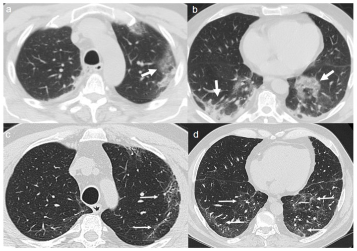
Figure 6.
(a,b) CT shows a “crazy paving pattern” peripherally located in the upper left lobe (arrow in a) and in the lower lobes (arrows in b). (c,d) CT after 4 months from the onset of symptoms shows persistence of mixed pattern characterized by interlobular septal thickening (thin arrows in c) and patchy GGOs (thin arrows in d).

