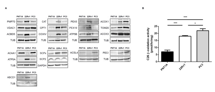
Figure 1.
Peroxisome and mitochondria protein and metabolic changes in prostate cancer (PCa). (A) Western blot analysis showing the expression levels of the peroxisomal proteins 70-kDa peroxisomal membrane protein (PMP70), acyl-CoA-binding domain-containing protein 5 (ACBD5), catalase (CAT), peroxin 5 (PEX5), peroxin 19 (PEX19), acyl-CoA oxidase 1 (ACOX1), acyl-CoA oxidase 3 (ACOX3), 3-ketoacyl-CoA thiolase (ACAA1), sterol carrier protein x (SCPx), acyl-CoA synthetase 4 (ACSL4), 3,2-trans-enoyl-CoA isomerase (PECI), and ATP binding cassette subfamily D member 1 (ABCD1), and the mitochondrial proteins voltage-dependent anion-selective channel protein 1 (VDAC1), carnitine palmitoyl transferase 1 (CPT1), cytochrome c oxidase subunit 4 (COXIV), ATP synthase subunit alpha and beta (ATP5A, ATP5B), mitochondrial import receptor subunit (TOM20), and sterol carrier protein 2 (SCP2), representative of at least three independent experiments. Tubulin (TUB) and glyceraldehyde 3-phosphate dehydrogenase (GAPDH) were used as loading controls. A densitometric quantification of the immunoblots is presented in Figure S1. (B) Cerotic acid (C26:0) peroxisomal β-oxidation activity in PNT1A, 22Rv1, and PC3 cells. Data represent means of three independent experiments and the bars represent standard error of the mean (SEM) of the mean. * p < 0.05 *** p < 0.001.

