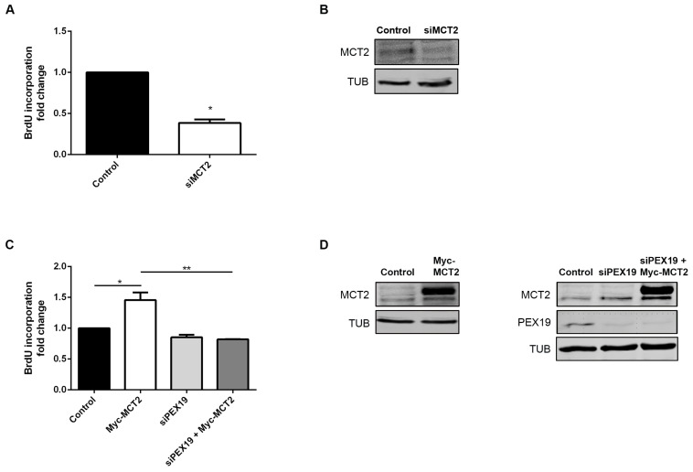
Figure 4.
MCT2 localization at the peroxisomal membranes is associated with PCa proliferation. (A) Effect of the silencing of MCT2 on 22Rv1 cell proliferation, measured by BrdU incorporation assay. Values are presented in fold change compared to control cells. Data represent means of three independent experiments and the bars represent SEM of the mean. * p < 0.05 ** p < 0.01 (B) Western blot analysis showing the expression levels of MCT2 and TUB in control and MCT2-silenced 22Rv1 cells. (C) Effect of Myc-MCT2 overexpression, silencing of Pex19, and MCT2 overexpression in the absence of PEX19 on 22Rv1 cell proliferation, measured by BrdU incorporation assay. Values are presented in fold change compared to control cells. Data represent means of three independent experiments and the bars represent SEM of the mean. * p < 0.05 ** p < 0.01 (D) Western blot analysis showing the expression levels of MCT2, PEX19, and TUB in control and MCT2-overexpressed and/or PEX19-silenced 22Rv1 cells. A densitometric quantification of the immunoblots is presented in Figure S1, since whole western blots are presented at Figure S5.

