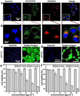
Figure 3.

In vitro PDT evaluation of fibrillar‐transformable PWG nanoparticles on MCF‐7 cells. a) Colocalization of PWG nanostructures in MCF‐7 cancer cells. CLSM images indicating the nuclei (stained with Hoechst), lysosomes (LysoTracker Green DND‐26), and PWG nanostructures (red), including the overlay. b) Magnified CLSM images of (a). The scale bar is the same for all images. c) CLSM images of intracellular 1O2 generation in MCF‐7 cells treated with PWG nanoparticles at pH 7.4. d) CLSM images of intracellular 1O2 generation in MCF‐7 cells treated with PWG nanoparticles at pH 6.5. e) Dark cytotoxicity and phototoxicity of PWG nanoparticles in MCF‐7 cells at pH 7.4 determined by MTT assay. f) Dark cytotoxicity and phototoxicity of PWG nanoparticles in MCF‐7 cells at pH 6.5 determined by MTT assay.
