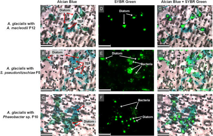
Fig. 1.

Micrographs of A. glacialis strain A3 co‐cultures with bacterial isolates. Diatom TEP were stained with alcian blue (A–C). Diatom and bacterial DNA was stained with SYBR Green I (D–F). Composite images show bacterial attachment mostly to TEP for S. pseudonitzschiae F5 and Phaeobacter sp. F10 but not A. macleodii F12 (G–I). Background in light micrographs show membrane filters. Scale bar represents 25 μm for all panels.
