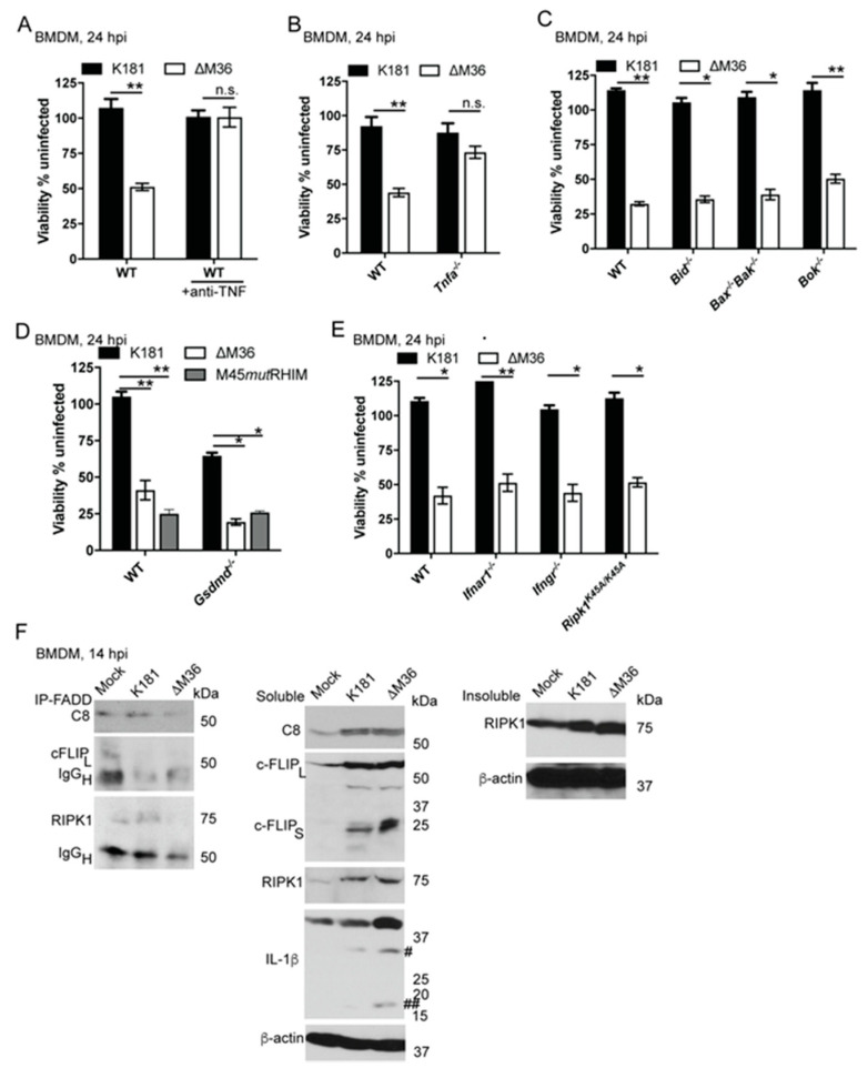
Figure 2.
Tumor necrosis factor (TNF) drives ∆M36-dependent apoptosis in BMDM. (A) Relative viability of WT BMDM infected with K181 or ∆M36 (MOI = 10) for 24 h in the absence or presence of neutralizing anti-TNF (100 μg/mL). anti-TNF was added in medium 1 hpi. (B–E) Relative viability of mutant BMDM when compared to WT cells at 24 hpi with indicated viruses. Viability graphs reflect data pooled from 3 to 5 independent experiments each containing 3 replicates. (F) IB showing the association of CASP8 (C8; 55 kDa), cellular FLICE inhibitory protein (cFLIP) (full length 55 kDa, indicated as cFLIPL) and RIPK1 (75 kDa) with Fas-associated via death domain (FADD) when Triton-X solubilized cell lysates from K181- or ∆M36-infected WT BMDM were subjected to immunoprecipitation at 14 hpi using anti-FADD antibody (left panel). Immunoglobulin (IgG) heavy chain (IgGH; 50 kDa) was detected when membranes were incubated with either anti-cFLIP or RIPK1 antibodies. Middle panel indicates total levels of C8, cFLIPL, as well as cFLIP short (cFLIPS; 25 kDa), RIPK1, and IL-1β with both total (#; 31 kDa) and processed forms (##; 17 kDa) were detected in the soluble cell lysates. Right panel indicates RIPK1 level in the detergent-insoluble fraction. β-actin (38.5 kDa) serves as loading control. IB data are representative of 3 independent experiments. Lines above bars indicate the two groups compared for significance values using paired, parametric Student’s t-test where * is <0.05, ** is <0.001, and n.s. represents nonsignificant.

