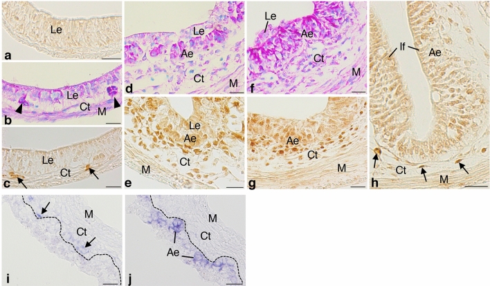
Figure 3.
Spatio-temporal expression of Foxl1 in the X. laevis intestine during metamorphosis. Cross sections were immunostained with anti-Foxl1 antibody (a,c,e,g,h) and stained with methyl green-pyronin (b,d,f), or hybridized with antisense Foxl1 (i) or c-Myc probes (j). (a) Stage 57. No cell is positive for Foxl1. (b,c) Stage 60. Adult stem/progenitor cells appear as islets strongly stained red with pyronin ((b), arrowheads) between the larval epithelium (Le) and the connective tissue (Ct). Some connective tissue cells become positive for Foxl1 ((c), arrows). (d,e) Stage 61. Both primordia of the adult epithelium (Ae) and the connective tissue increase in cell number (d). Connective tissue cells positive for Foxl1 become numerous close to the adult epithelial primordia (e). (f,g) Stage 62. The adult epithelium mostly replaces the larval one (f). Most of the connective tissue cells surrounding the adult epithelium are positive for Foxl1 (g). (h) Stage 66 (end of metamorphosis). Connective tissue cells positive for Foxl1 (arrows) are small in number and tend to be localized in the trough of newly-formed intestinal folds (If). (i,j) Stage 62. Foxl1 mRNA (i, arrows) is detected as dark blue deposits in the connective tissue cells close to adult epithelial primordia expressing c-Myc (j). The dashed-lines indicate the boundary of the epithelium and connective tissue. M; muscles. Bars, 20 μm.

