
Figure 2. Inflammatory cell infiltration of lacrimal glands of male NOD mice at and after 6 weeks of age.
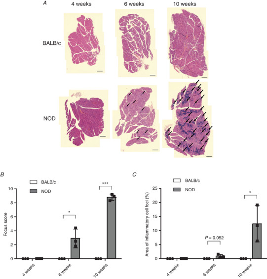
A, representative cross‐sections of haematoxylin and eosin‐stained lacrimal glands from male BALB/c and NOD mice at 4, 6 and 10 weeks of age. Arrows show infiltration of inflammatory cells. Scale bars represent 0.5 mm. B and C, the focus score (B) and the area of inflammatory cell foci (C) were determined in the lacrimal glands of male BALB/c and NOD mice at 4, 6 and 10 weeks of age. The obtained values are presented as the mean ± SD: * p < 0.05, ** p < 0.01, *** p < 0.001 versus BALB/c group. All experiments were conducted with three animals in each group.