Figure 3.
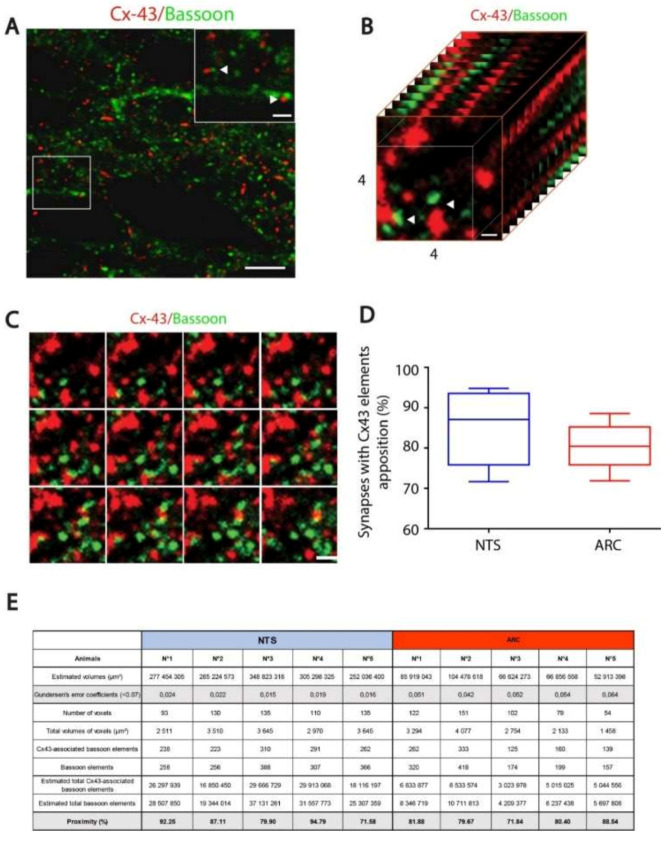
(A): Confocal images of connexin-43 (Cx43) (red) and bassoon (green) double labeling immunohistochemistry performed within the nucleus tractus solitarius (NTS). Scale bar: 5 μm. Inset: Arrows show Cx43 labelling in close vicinity of synapses labelled by presynaptic bassoon protein. Scale bar 1 µm. (B): An example of a voxel (4 × 4 µm) used for the quantification of Cx43/bassoon appositions. Scale bar: 0.5 µm. (C): Twelve z-stack images defining the voxel illustrated in B and allowing the identification of Cx43/bassoon appositions. (D): Stereological estimation of Cx43/bassoon appositions in the NTS (85.13%) and arcuate nucleus of the hypothalamus (ARC) (80.46%) nuclei. (E): Table gathering the different stereological values acquired or estimated for each studied animal.

