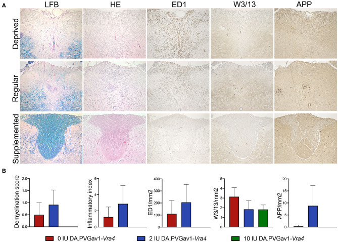
Figure 3.
CNS inflammation and demyelination in the congenic rats upon different concentration of vitamin D in the diet. (A) Images of the spinal cord cross-sections of DA.PVGav1-Vra4 rats subjected to different levels of vitamin D in their diet. Histopathological and IHC analyses were performed on an average of 18 paraffin embedded spinal cord cross-sections per animal harvested on day 33 p.i. (n = 4- 6 per diet group). The sections were stained with LFB, HE, against ED1, W3/13, and APP, respectively, in order to assess: (B) demyelination score (DM), inflammatory index (I.I.), recruitment of ED1+ microglia/macrophages, W3/13+ lymphocytes, and axonal damage, respectively. Statistics were calculated using one-way ANOVA with Tukey correction for multiple testing.

