Figure 1.
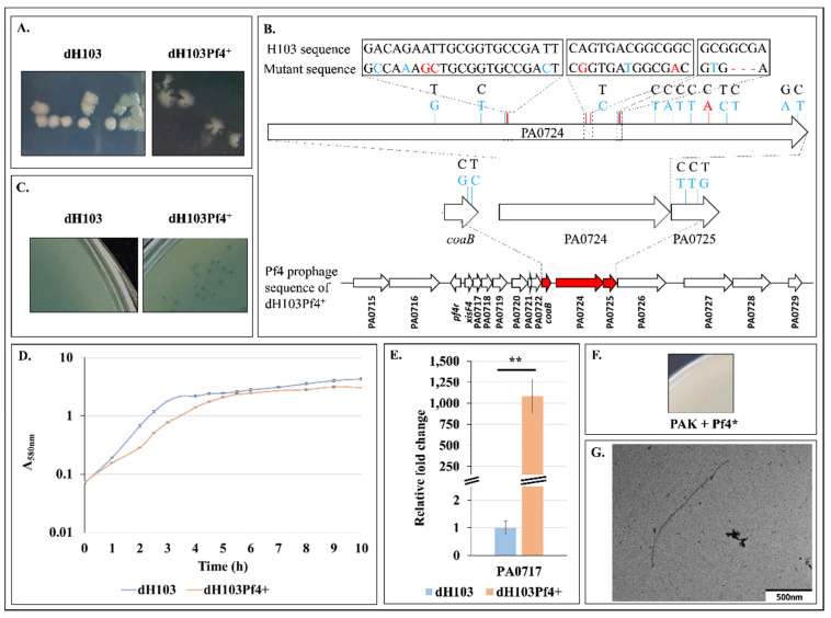
Isolation and description of a Pf4 phage variant. (A). Colony morphology of P. aeruginosa dH103 and dH103Pf4+ strains. (B). Gene mapping of the coaB-PA0725 Pf4 prophage region in dH103Pf4+ with mutations compared to H103. Silent mutations are represented in blue. Mutations and deletion are represented in red. (C). Lysis plaque assay using supernatant of dH103 and dH103Pf4+ performed on H103 strain. (D). Growth curves of dH103 and dH103Pf4+ ± SEM. (E). Relative mRNA levels of PA0717 in dH103 (blue bar) and dH103Pf4+ (orange bar) ± SEM as determined by RT-qPCR experiments. (F). Lysis plaque assay using supernatant of dH103Pf4+ performed on PAK strain. (G). TEM picture of Pf4* phage. Lysis plaques, growth and RT-qPCR experiments were assayed at least four times independently. Statistics were achieved by unpaired (two samples) two-tailed t-test. ** p < 0.01.

