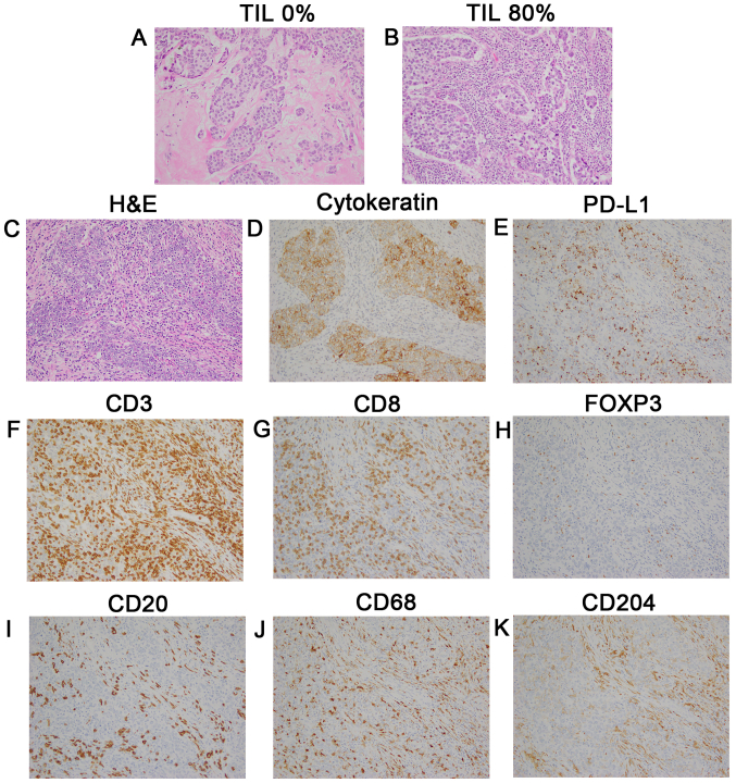
Figure 1.
Representative images of TILs and immune subsets evaluated by IHC staining. Magnification, ×200. (A) TILs, 0%; and (B) TILs, 80%. TMA samples of a triple-negative tumor (stage II; HG 3; Ki-67 80%) according to (C) H&E staining, (D) cytokeratin, (E) PD-L1 expression (3+ on TCs and 2+ on ICs), (F) CD3+, (G) CD8+, (H) FOXP3+, (I) CD20+, (J) CD68+ macrophages, (K) CD204+ macrophages. TILs, tumor-infiltrating lymphocytes; IHC, immunohistochemical; TCs, tumor cells; ICs, immune cells.

