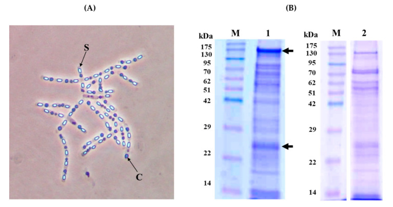
Figure 1.
Crystal morphology and SDS-PAGE analysis of the KhF strain. (Panel A) Phase-contrast microscopy (×100 magnification). Letters “S” and “C” point to spore and crystal, respectively. (Panel B) SDS-PAGE analysis. Lane 1; Spore/crystal mixture, lane 2; partially solubilized KhF crystals in 50 mM carbonate buffer (pH 10.5). Lanes M; molecular mass marker (Pink pre-stained protein ladder, Nippon genetics). The arrows point to the major bands of 130 and 23 kDa.

