Figure 3.
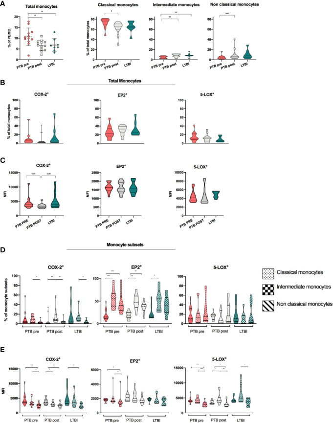
Expression of monocytes, monocyte subsets and eicosanoid expression in stages of Mtb infection. (A) Distribution and frequencies of monocytes and monocyte subsets in different clinical stages of Mtb infection. PTB at time of diagnosis (PTB pre, pink, N = 13), PTB at end- of- treatment (PTB post, grey, N = 13), and LTBI (green, n = 8). Monocyte subsets were defined as classical monocytes (CD14++CD16-), Intermediate monocytes (CD14++CD16+) and non-classical monocytes (CD14+CD16++) (Gating strategy Supplementary Figure 1A ) (29). (B) Frequencies of COX-2, EP2, and 5-LOX in unstimulated samples in total monocytes. (C) Mean fluorescence intensity (MFI) of COX-2, EP2, and 5-LOX in unstimulated samples in total monocytes. Frequencies (D) and MFI (E) of COX-2, EP2, and 5-LOX in unstimulated classical, intermediate, and non-classical monocyte subsets, presented by fill pattern, stratified by patient group (color). Mann-Whitney test was used for unpaired samples, Wilcoxon signed rank test used for paired samples. Statistical significance represented by asterisk: *p < 0.05; **p < 0.01; ***p < 0.001.

