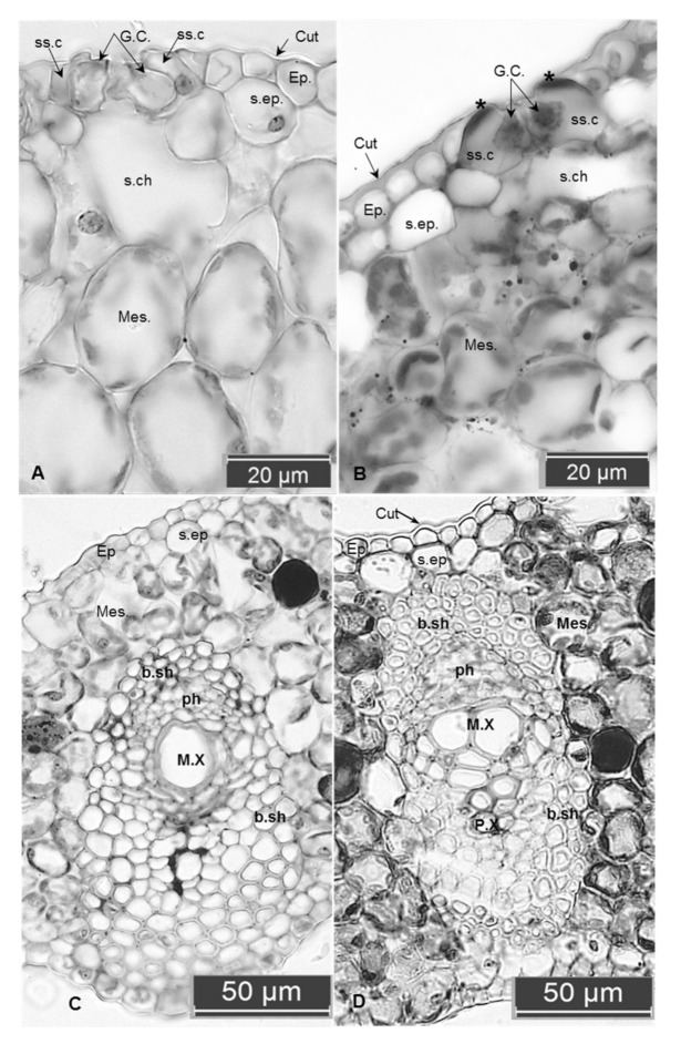
Figure 1.
Cross-section of in vitro leaves of a date palm: (A,C) control untreated leaves, (B,D) treated with 20 g L−1 PEG. (A) The thin cuticle layer, larger mesophyll cells with fewer chloroplasts, greater substomatal chamber and intercellular spaces. Note the opened stomata and the same thickness of both ventral and dorsal walls of the guard cells, thin-walled of subsidiary cells. (B) The thick cuticle, compacted mesophyll cells with smaller substomatal chambers. Note the closed stomata, increasing in the thickness of ventral walls of the guard cell, and also the outer tangential walls of the two subsidiary cells appear lignified (the two stars). (C) Weak differentiation of the vascular bundle sheath compared to the well-developed one appears as lignified cells around phloem and xylem in (D). Abbreviations: Cut. cuticle; Ep. epidermis; GC. guard cells; ss.c. subsidiary cells; s. ep. sub epidermal layer; s.ch. substomatal chamber’ mes. mesophyll; b.sh. bundle sheath; ph. phloem; M.X. meta xylem; P.X. proto xylem.

