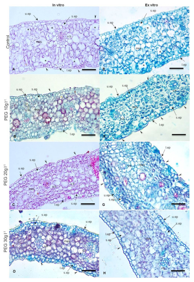
Figure 3.
Cross sections in in vitro date palm leaves (cv. Sewi) grown in media supplemented with different levels of PEG (A–D) and after 4 weeks from ex vitro transplantation (E–H). (A) Abnormal structures appeared as thin cuticle, large mesophyll cells and fewer chloroplasts, large intercellular spaces, and substomatal chambers. (B,C) Organized mesophyll with reduction in intercellular spaces and well developed cuticle layer. (D) Compacted mesophyll, thick cuticle. (E) Leaves still have thin cuticle, loose mesophyll, but the plastids appear in higher density than observed in (A,F). More organized mesophyll, closed stomata. (G) Well developed compacted mesophyll and slightly sunken stomata. (H) Thick cuticle, closed stomata. Abbreviations: u.ep. upper epidermis; s.ep. subepdermal layer; l.ep. lower epidermis; mes. Mesophyll; v.b. vascular bundle. Arrowheads point to stomata, stars point to substomatal chambers and intercellular spaces. Bar = 200 µm.

