Fig. 4.
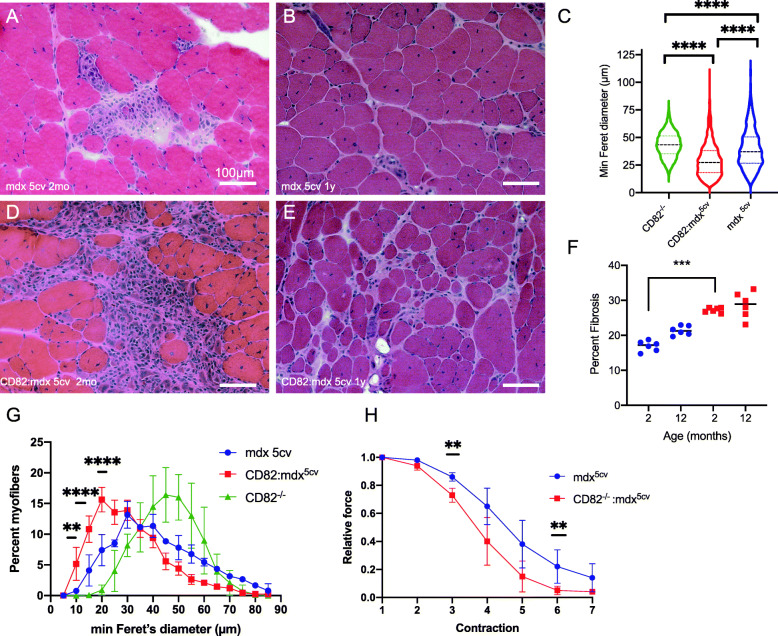
Ablation of CD82 in dystrophic muscle leads to worsened skeletal muscle phenotype. H&E images of the quadriceps muscles from mdx5cv (a, b) and from CD82−/−:mdx5cv mice (d, e). Images were taken from tissues at 2 months (a, d) and 1 year (b, e) of age. c Comparisons of myofiber size using ANOVA shows significantly smaller myofibers in both strains of dystrophic mice compared to CD82−/− mice, with CD82−/−:mdx5cv myofibers being the smallest. f Quantification of fibrosis at 2 months and 1 year of age shows increased presence of fibrotic tissue in young CD82−/−:mdx5cv compared to control mdx5cv mice. Examples of images used in quantification are shown in Supplementary Figure 4F, G. g Distribution plots of myofiber size show significantly smaller myofibers in CD82−/−:mdx5cv compared to control mdx5cv and CD82−/− muscles. h Isometric force analysis following serial eccentric contractions demonstrated significantly decreased muscle force production after each contraction in CD82−/−:mdx5cv compared to control mdx5cv mice. N = 6/group (**p ≤ 0.01; ***p < 0.001; ****p < 0.0001). Scale bars: 100 μm

