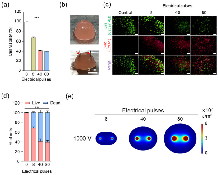
Figure 2.
In vitro cellular experiments to establish electrical conditions for irreversible electroporation (IRE). (a) In vitro cytotoxicity of LLC under various electrical pulses in IRE (voltage: 1000 V). Asterisks indicate statistical significance compared to the control group (*** p < 0.0001, n = 3). (b) Digital photograph of the 3D tumor model. LLC cells were cultured in a hydrogel containing alginate and gelatin, and a needle electrode with a 3 mm spacing was used for the IRE. The red arrow indicates the electrode. (c) In vitro live and dead cell viability assay of LLC under various electrical pulses in the IRE (at 1000 V). Live and dead cells were stained with Calcein AM (green) and propidium iodide (red), respectively (scale bars = 200 μm). (d) Quantitative analysis of the live and dead cell assay results. Asterisks indicate statistical significance compared to the control group (*** p < 0.0001, n = 3). (e) Finite element method (FEM) simulation images of IRE area. The color indicates the electric energy density level of the IRE zone with 8, 4, and 80 pulses (voltage: 1000 V and pulse duration: 100 μs).

