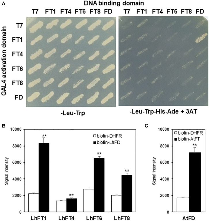
FIGURE 7.
Protein–protein interactions between the LhFT proteins and LhFD. (A) The LhFT1, LhFT4, LhFT6, LhFT8, and LhFD proteins were fused to the GAL4 DNA-binding domain (BK) or GAL4 activation domain (AD). pGBKT7 and pGADT7 were used as the negative controls, for bait and prey, respectively. Yeast cells were grown on double-selection medium (−Leu, and −Trp; left) and quadruple-dropout medium (−Leu, −Trp, −His, and −Ade) supplemented with 15 mM 3-AT (right) at 30°C for 3 days. (B) in vitro protein–protein interaction assay by AlphaScreen. AGIA-tagged LhFTs were incubated with biotinylated LhFD. The interaction intensity between LhFTs and LhFD was analyzed by AlphaScreen Biotinylated dihydrofolate reductase (DHFR), and E. coli was used as negative control. Data are mean ± SD. of three independent experiments (n = 3). **indicates the significant difference with student’s t-test (P < 0.01). (C) AlphaScreen assay for Arabidopsis FT–FD interaction. AGIA-tagged AtFD was incubated with biotinylated AtFT. Data are represented as mean ± SD of three independent experiments (n = 3).

