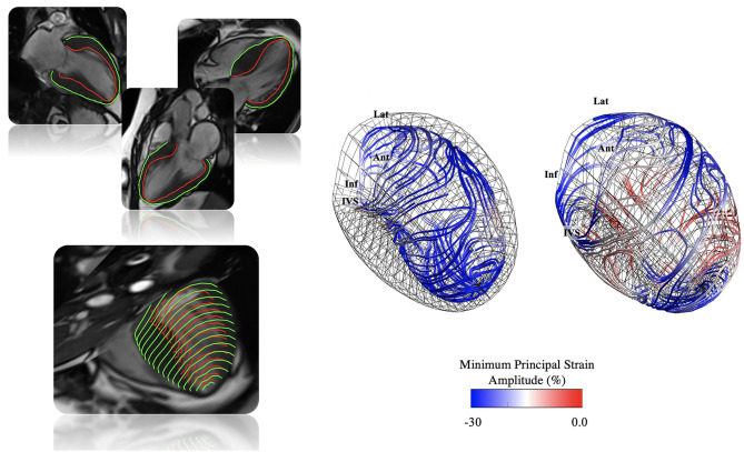
Figure 1.
Illustration of 3D-MDA workflow. Top left: Endocardial and epicardial contour tracing performed at end-diastole on long-axis cine images with automated registration to short-axis images and 3D mesh generation. Bottom left: A 4D displacement field is generated (1, 4, 6–8, 15), and used to deform the end-diastolic phase 3D LV mesh (1, 3, 4, 6) throughout the cardiac cycle (1, 4, 6). Right: From this dynamic mesh model strain, conventional and principal strain quantification is performed (16, 17). In the example, principal strain amplitude and direction lines are shown for a patient with confirmed hypertrophic cardiomyopathy (HCM).

