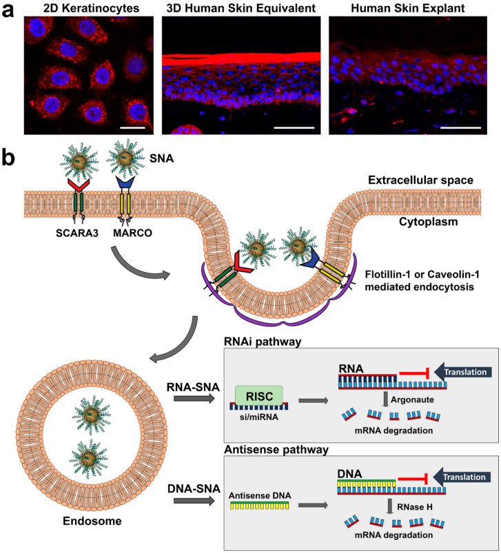
Figure 2.
Mechanism of SNA uptake in keratinocytes. (a) Confocal imaging of keratinocyte uptake of fluorescently (Cy5) labeled SNAs (red) in 2D, 3D and human explant culture; scale bars in (a) from left to right: 20 mm, 50 mm, 50 mm. (b) SNAs are: (i) first detected on the keratinocyte cell surface by class A scavenger receptors SCARA3 and MARCO; (ii) taken up primarily by flotillin-1-mediated endocytosis, but also caveolin-1-mediated endocytosis; (iii) deposited into endosomes; and (iv) released from endosomes to suppress gene expression via the RNAi (RNA-SNAs) or antisense (DNA-SNA) pathway; in the RNAi pathway mRNA translation is blocked by antisense RNA or target mRNA is degraded by Argonaute of the RISC complex, whereas in the antisense pathway antisense DNA can block translation or initiate mRNA degradation by RNase H.

