Figure 3.
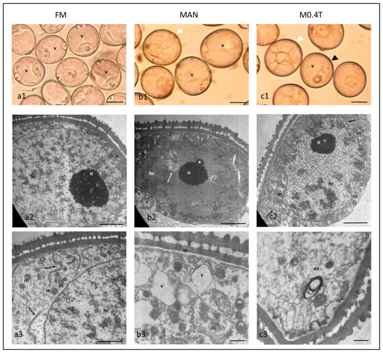
Morphological and ultrastructural characterization of freshly isolated microspores of bread wheat cv. Pavon (FM) (a1–a3), after 0.7 M mannitol (MAN) (b1–b3) and 0.7 M mannitol with 0.4 µM TSA treatments (M0.4T) (c1–c3). FM showed a large vacuole (v), a euchromatin nuclei with small patches of heterochromatin (hc), and a prominent nucleolus (a1–a2); cytoplasm with abundant free ribosomes, mitochondria (m), and RER (black arrows) (a3). Both MAN and M0.4T showed larger microspores, some with SLM morphology (b1, c1, white arrowheads) or symmetrically divided (c1, black arrowhead); a euchromatic nucleus with patches of heterochromatin (hc) (b2, c2), a small nucleolus with a Cajal body (asterisk) (b2), and invaginations of the nuclear envelope (b2, white arrow); cytoplasm with vacuoles (v), free ribosomes, mitochondria (m), and autophagosome (au) (b2, b3, c3); and an intine layer (in) (b3, c3). Scale bars for a1–c1= 20 µm; scale bars for a2, b2, c2 = 2 µm; scale bars for a3 = 1 µm; and scale bars for b3, c3 = 0.5 µm.

