
Figure 3.
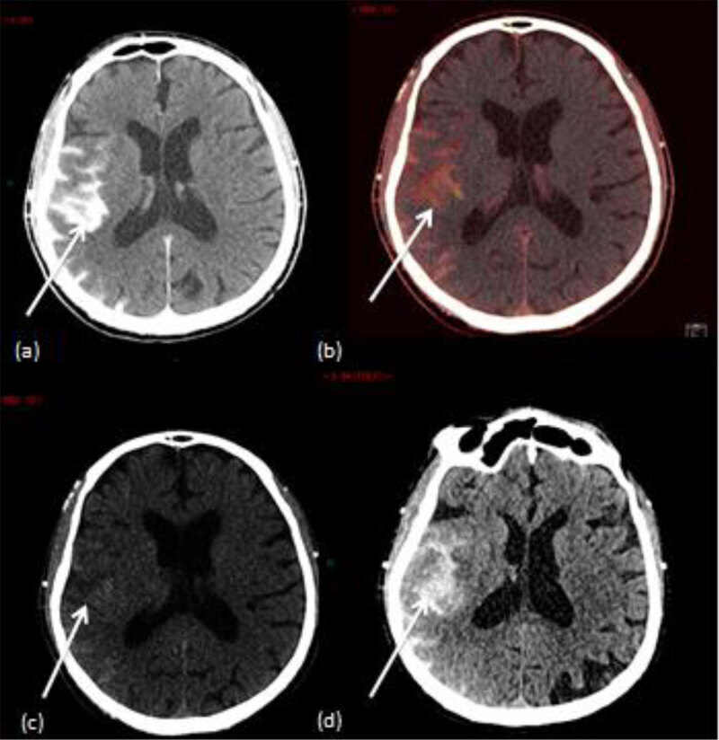
A case of a 77 years old patient who underwent intra-arterial thrombectomy (right middle cerebral artery). (a) Mixed DECT image shows an hyperattenuation in the perisylvian cortex. (b) IOM shows contrast material staining in the perisylvian cortex. (c) VNC image shows a subtle hyperattenuation in the perisylvian cortex. (d) Follow-up conventional brain CT shows a persistant hyperattenuation in the perisylvian cortex which confirms brain hemorrhage.