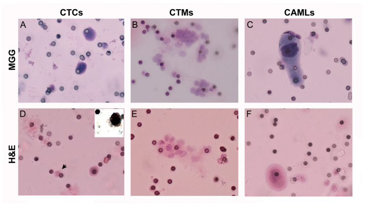
Figure 1.
Representative staining with hematoxylin and eosin (H&E) and May–Grünwald–Giemsa (MGG) of circulating cells enriched by ScreenCell filters from blood samples of ACC patients. Circulating tumor cells (CTCs, A,D), circulating tumor microemboli (CTMs, B,E), and cancer associated macrophages (CAMLs, C,F) were separated by ScreenCell devices from blood samples of ACC patients (magnification: 40×) and stained with hematoxylin and eosin (H&E) or May-Grünwald Giemsa (MGG). A polymorphonuclear leukocyte with the typical multilobated nuclei is indicated by the arrow (D) for comparison with a CTC. The small pores of 6 µm are visible as small dots non homogenously distributed on the filter membrane. Inset in panel (D) shows the nuclear positivity to steroidogenic factor-1 (SF-1) antibody in a representative CTC obtained in a parallel filter from the same blood sample.

