Figure 5.
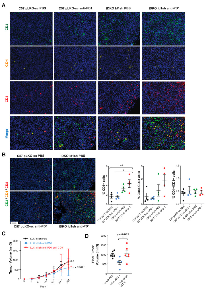
CD8+ T cells inflammatory infiltrate may mediate the anti-tumor activity observed after the Id1 and PD-1 blockade. (A) Quantitative multiplex IHC (CD3, CD4, CD8 and multiplex) in representative sections of tumors belonging to group of mice with extreme phenotypes [Id1+/+/DPBS (pLKO-sc LLC), Id1+/+/anti-PD-1 (pLKO-sc LLC), Id1-/-/DPBS (Id1sh LLC) and Id1-/-/anti-PD-1 (Id1sh LLC); four mice per group]. (B) Representative merged images derived from the multiplex quantification of CD3/CD4/CD8 in the same tumors as in A confirm the higher TIL when tumors lack Id1 and are treated with anti-PD-1 therapy. Quantification of the percentage of CD3, CD8/CD3, and CD4/CD3 positive cells. (C) In vivo tumor growth of Id1sh-LLC cells in C57BL/6J mice treated with CD8+ T cells depleting antibodies. Tumors were measured on days 7, 10, 14, 17, 21, and 24. (D) Final tumor volumes of the same three mice groups as in C at day 24 post-tumor injection (Id1sh/anti-PD-1/anti-CD8: 962.1 [786.0–1360] mm3; Id1sh/DPBS: 914.5 [853.6–1160] mm3, n.s.; Id1sh/anti-PD-1: 602.3 [386.4–740.5] mm3, p = 0.0425). The data are reported as the median with the interquartile range or mean ± SD. * p < 0.05, ** p < 0.01, *** p < 0.001, n.s. (not significant). Scale bar, 30 µm.

