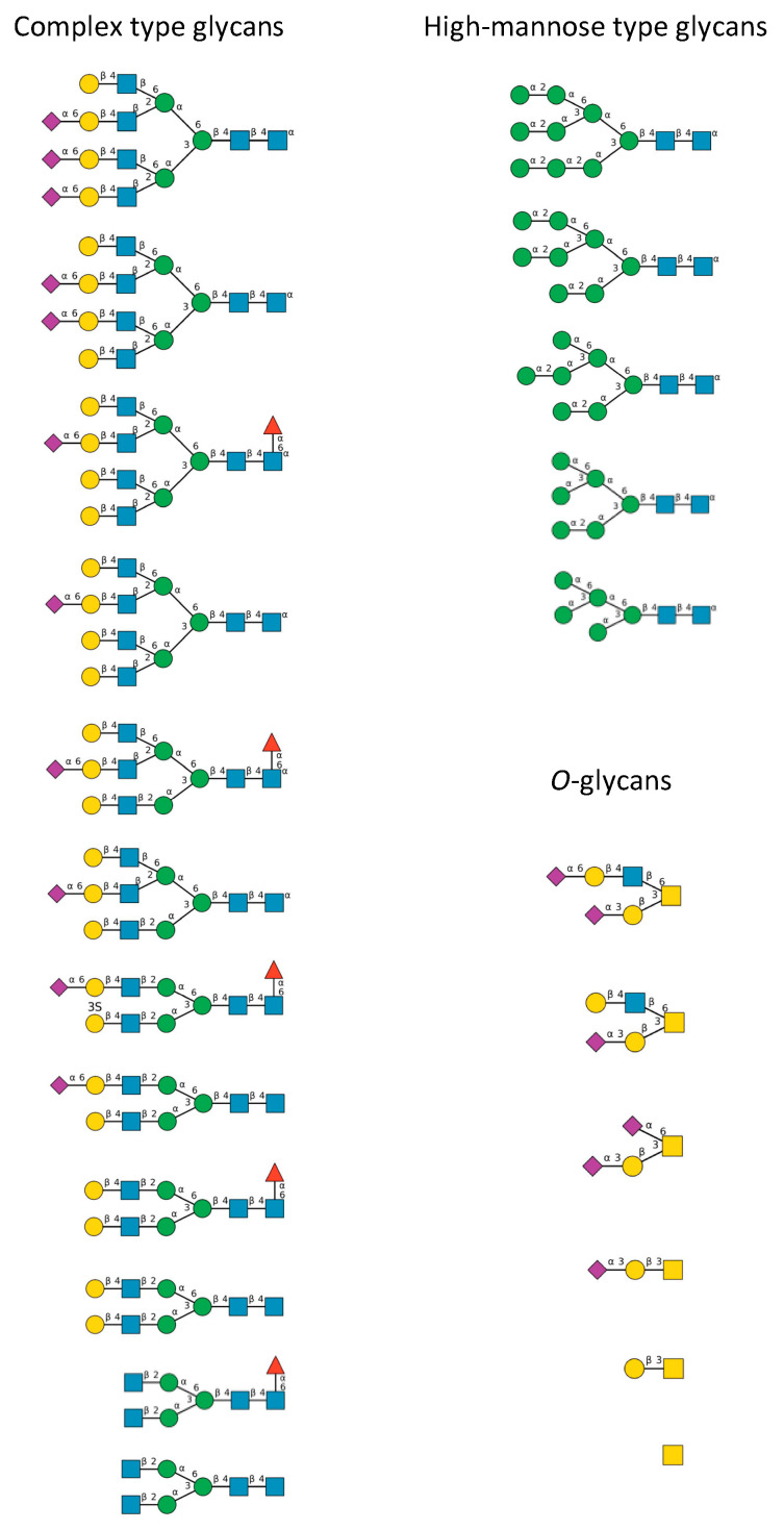
Figure 10.
Diversity of the N-glycans of the biantennary complex type (left frame) and high-mannose type (upper right frame), and O-glycans (lower right frame), identified in the S-glycoprotein forming the spikes at the surface of the SARS-CoV-2 envelope [26]. Symbols used to represent the N- and O-glycans: blue squares: N-acetylglucosamine (GlcNAc), green circles: mannose (Man), yellow circles: galactose (Gal), red triangle: fucose (Fuc), purple diamonds: sialic acid (Neu5Ac), yellow square: N-acetylgalactosamine (GalNAc).

