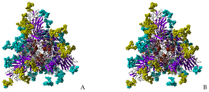
Figure 14.
Glycosylation of trimeric S-glycoprotein of SARS-CoV-2. (A) Front view of the trimeric S-glycoprotein of SARS-CoV-2 showing the high-mannose type glycans (colored yellow) specifically recognized by Man-specific lectins KAA-2 and HRL-40 from the red algae Kappaphycus alvarezii [10,13] and Halimeda renschii [14], and OAA from the blue-green alga (cyanobacterium) Oscillatoria agarddhii [16]. Other complex N-glycans decorating the monomer weakly or not recognized by the lectins, are colored cyan. (B) Front view of the trimeric S-glycoprotein of SARS-CoV-2 showing the high-mannose type glycans (colored yellow) specifically recognized by the Manα1,2-specific lectin BCA from the green alga Boodlea coacta [11]. Other complex N-glycans decorating the monomer weakly or not recognized by BCA, are colored cyan.

